Ficheiro:Phycoerythrin 545 1XG0.gif
Phycoerythrin_545_1XG0.gif (400 × 474 píxeles; tamaño do ficheiro: 85 kB; tipo MIME: image/gif)
| Este ficheiro procede de Wikimedia Commons. A continuación móstrase a información da súa páxina de descrición. Commons é un repositorio libre de ficheiros multimedia. Pode contribuír alí cargando as súas imaxes. |
Resumo
| Descrición |
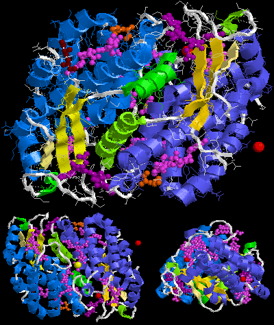
English: Crystal structure of Phycoerythrin 545 from Rhodomonas CS24 (PDB ID: 1XG0). This image was created with RasTop (Molecular Visualization Software) and Adobe Photoshop Elements. |
| Data | |
| Orixe |
Obra própria, usando: the Protein Data Bank (PDB) structural data: |
| Autoría | Darekk2 using the cited above Protein Data Bank (PDB) structural data |
Licenza
Attribution: The author of the work, the original authors of the Protein Data Bank (PDB) structural data and the molecular graphics program used. PDB data and software citation example:
Image of PDB (Protein Data Bank) ID 1XG0 (Doust, A.B., Marai, C.N.J., Harrop, S.J., Wilk, K.E., Curmi, P.M.G., Scholes, G.D. (2004) Developing a structure-function model for the cryptophyte phycoerythrin 545 using ultrahigh resolution crystallography and ultrafast laser spectroscopy. J.Mol.Biol., 344: 135-153) created with RasTop (Molecular Visualization Software).
The RCSB Protein Data Bank (PDB) states on its website in the Policies & References section:
http://www.rcsb.org/pdb/static.do?p=general_information/about_pdb/policies_references.html archive copy at the Wayback Machine
Data files contained in the PDB archive (ftp://ftp.wwpdb.org) are free of all copyright restrictions and made fully and freely available for both non-commercial and commercial use. Users of the data should attribute the original authors of that structural data. (...) Images created using PDB data and other software should cite the PDB ID and the molecular graphics program used.
the original authors of the Protein Data Bank (PDB) structural data
and the molecular graphics program used
- Vostede é libre de:
- compartir – copiar, distribuír e difundir a obra
- facer obras derivadas – adaptar a obra
- Baixo as seguintes condicións:
- recoñecemento – Debe indicar a debida atribución de autoría, fornecer unha ligazón á licenza e indicar se se realizaron cambios. Pode facer isto de calquera forma razoable, mais non nunha forma que indique que quen posúe a licenza apoia ou subscribe o seu uso da obra.
- compartir igual – Se altera, transforma ou amplía este contido, debe publicar as súas contribucións baixo a mesma licenza ou outra compatible á orixinal.
Historial do ficheiro
Prema nunha data/hora para ver o ficheiro tal e como estaba nese momento.
| Data/Hora | Miniatura | Dimensións | Usuario | Comentario | |
|---|---|---|---|---|---|
| actual | 11 de outubro de 2012 ás 15:52 | | 400 × 474 (85 kB) | Darekk2 | User created page with UploadWizard |
Uso do ficheiro
A seguinte páxina usa este ficheiro:
Uso global do ficheiro
Os seguintes wikis empregan esta imaxe:
- Uso en en.wikipedia.org
- Uso en it.wikipedia.org